


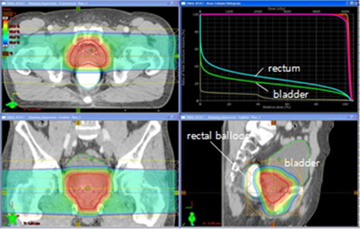
- Proton therapy is suitable for prostate cancer because it minimizes the radiation dose delivered to healthy rectal and bladder tissue
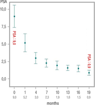
- Prostate-specific antigen is known to decrease after proton therapy.
- At 2 years old, a child was diagnosed with ependymoma. She was treated with proton therapy between November 2009 and January 2010 after undergoing surgery and chemotherapy.

- At 6 years old, she has had no evidence of disease for 3 years and 2 months. Although she has a little difficulty in balancing when walking and going up the stairs, her speech and vocabulary are close to the levels of other children of her age.
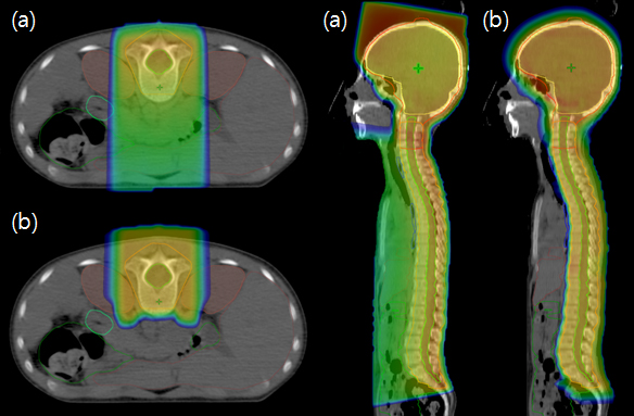
- Proton therapy can be used to treat tumors in the craniospinal space and minimizes the radiation dose delivered to the rest of the body. (a) X-ray and (b) proton therapy.
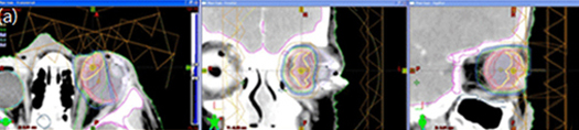
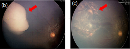
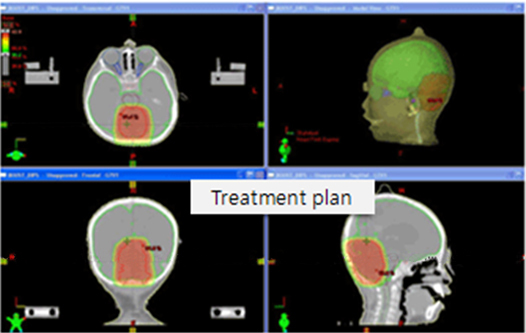
- A 3-year-old boy with retinoblastoma received proton therapy. The optic nerve, lens, normal retina, lacrimal gland, and orbital bone received minimal doses of radiation. He is in his 4th year of elementary school, and has shown no evidence of disease 4 years and 9 months after treatment.
His vision is also good. (a) Proton therapy plan. (b, c) Ophthalmoscopy before (b) and 1 month after (c) proton therapy (necrotic cavity).
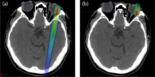
- Proton therapy can be used to treat uveal melanoma and minimizes the radiation dose delivered to the brain. (a) X-ray and (b) proton therapy.
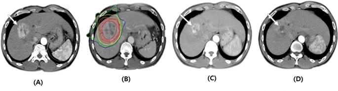
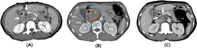
Complete response (CR) of a primary tumor to proton beam therapy (PBT).
- (A) Pretreatment CT scans showing the primary tumor (arrow).
- (B) The patient underwent PBT.
- (C) CT scans at 3 months after PBT demonstrating shrinkage of the primary tumor (arrow).
- (D) CT scans at 8 months after PBT demonstrating complete regression of the primary tumor (arrow).
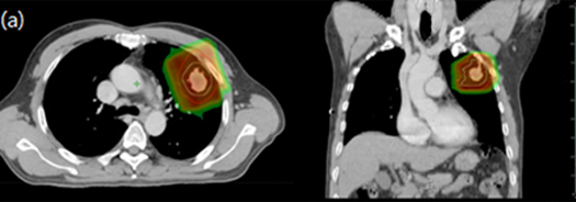
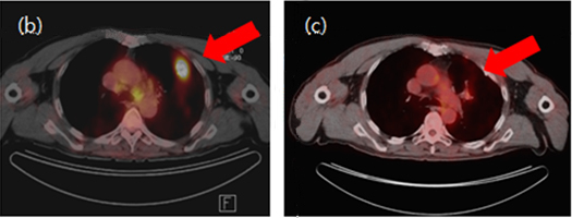
- A 56-year-old male with stage I non-small cell lung cancer was contraindicated for surgery because of poor lung function. Therefore, he received proton therapy instead. He had no evidence of disease 4 years after treatment. (a) Proton therapy plan. (b, c) Before (b) and 4 years after (c) proton therapy
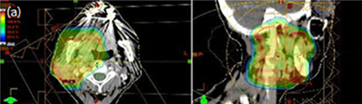
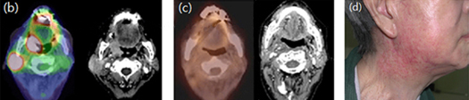
- An 84-year-old female with locally advanced oropharyngeal cancer received proton therapy. The contralateral parotid gland received a minimal radiation dose. She had no evidence of disease 9 months after treatment. (a) Proton therapy plan. (b, c) Before (b) and after (c) proton therapy. (d) Skin changes following administration of 64.8 Gy in 27 fractions.
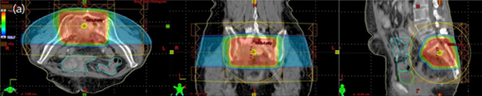
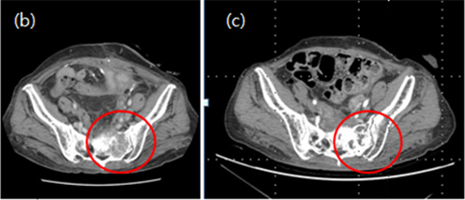
- A 72-year-old female was diagnosed with recurrent S-colon cancer in the sacrum, approximately 10 years after initial diagnosis. She was treated with proton therapy (72 Gy in 30 fractions). (a) Proton therapy plan. (b, c) Before (b) and 1 year after (c) proton therapy.
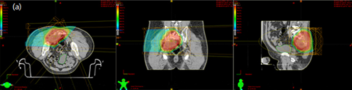
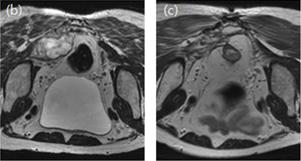
- A 62-year-old male with sacral chordoma received proton therapy (74.4 Gy in 31 fractions). The rectum and bowel received minimal radiation doses. (a) Proton therapy plan. (b, c) Before (b) and 4 years after (c) proton therapy.
Pancreatic Cancer?
Pancreatic cancer is a disease in which malignant cells form in the tissues of the pancreas. The pancreas produces digestive juices and hormones that regulate blood sugar.
The pancreas is located in the mid-abdominal area
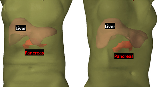 It is surrounded by several critical organs including the liver, kidneys, stomach and the small and large intestines.
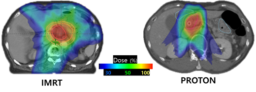
Why Proton?
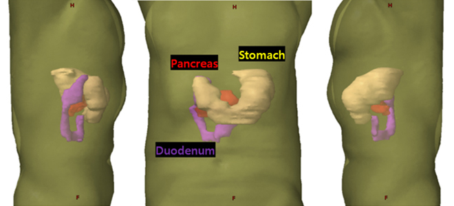
The use of traditional radiation therapy can be challenging, because of the pancreas’ proximity to nearby critical tissues and organs, including the small intestine, duodenum, stomach and spinal cord.
Proton therapy provides an opportunity for treatment that is less likely to compromise these surrounding tissues and organs. s
Clinical Case
- (A) pretreatment CT scans showing the primary tumor (arrow)
- (B) the patient underwent SIB-PBT
- (C) CT scans 3 months after SIB-PBT demonstrating Partial response(PR) of the primary tumor (arrow).